Biomineralization is a fascinating natural process by which living organisms produce hierarchical mineral structures with diverse functions. The “secrets” of biomineralization are explored by the scientists since decades but there are still open questions regarding its function, the regulating mechanisms and why and when biomineralization started.This process occurs through self-organization of organic and inorganic molecules and requires specific ambient conditions. It results in highly structured materials with remarkable physical and chemical properties. Examples are the formation of silicates in algae and diatoms, carbonates in invertebrates and phytoplanktonic algae, and calcium phosphates and carbonates in the hard tissues of vertebrates. The crescent number of works dealing with biomineralization highlights the interest of the scientific community which aims at understanding why, under specific circumstances, the process fails. Here I propose a selection of the latest papers published on this topic.
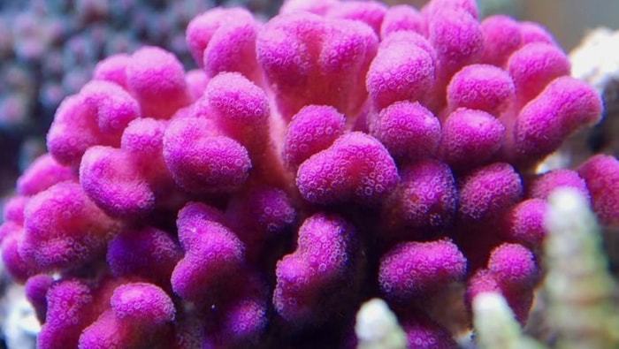
Coronado et al. (2019) published on Nature Communications the work titled “Impact of ocean acidification on crystallographic vital effect of the coral skeleton” where they have assessed crystallographic parameters of bio-aragonite to study the response of the reefbuilding coral Stylophora pistillata to experimental seawater acidification.

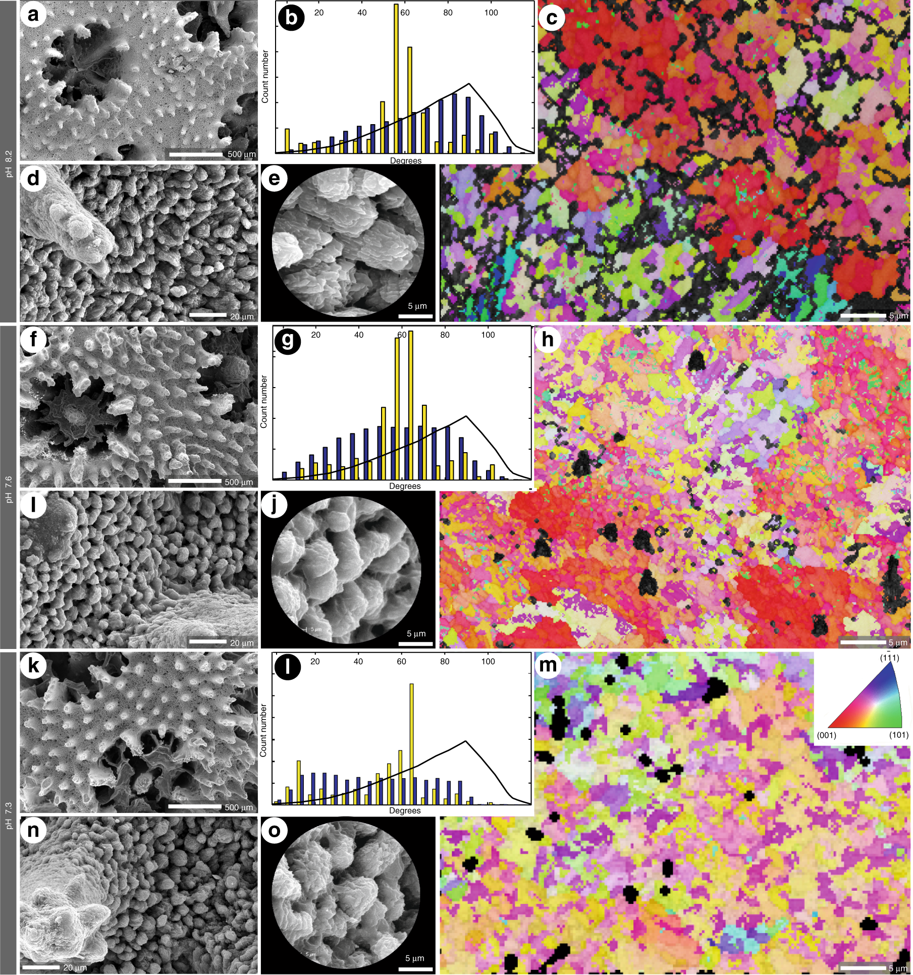
Structural features of Stylophora pistillata cultured at different pH conditions (from Coronado et al. 2019)
Skeletons formed under high pCO2 conditions show systematic crystallographic changes such as better constrained crystal orientation and anisotropic distortions of bio-aragonite lattice parameters due to increased amount of intracrystalline organic matrix and water content.
In the work titled “Terebratulide brachiopod shell biomineralization by mantle epithelial cells” by Roda et al (2019) published in the Journal of Structural Biology the authors wanted to focus on an unknown process in brachiopods: how the mineral is transported from outer mantle epithelium cells to the site of mineralization. So they imaged with TEM and FE-SEM ultrastructural characteristics of outer mantle epithelium cells on Magellania venosa shells to investigate the mineral transport pathways for shell secretion and to assess differences in cellular activity during mineralization.

Tang et al (2019) published in Nature Communication “Spiculogenesis and biomineralization in early sponge animals” where they report an early Cambrian sponge that had weakly biomineralized and hexactine-based siliceous spicules with large axial filaments and high organic proportions. This material, along with Ediacaran microfossils containing putative nonbiomineralized axial filaments, suggests that Precambrian sponges may have had weakly biomineralized spicules or lacked them altogether, hence their poor record. This work provides a new search image for Precambrian sponge fossils, which are critical to resolving the origin of sponge spiculogenesis and biomineralization.

Preservation of organic and biosilica structures in Vasispongia delicata (from Tang et al., 2019)
The recipe for making shells, spines, and coral skeletons is not only the same across many modern animal lineages, but is ancient—dating back 550 million years—and evolved independently more than once. This is what is presented in”Biomineralization by particle attachment in early animals ” by Pupa et al (2019) published in PNAS . The authors show that when echinoderms, mollusks, and cnidarians started biomineralizing in the Cambrian (more than 500 million years ago) these three phyla started doing it in precisely the same way: using attachment of amorphous nanoparticles.”
Modern and fossil nacre from 3 molluscan classes, exhibiting irregularly shaped nanoparticles (from Pupa et al. 2019)
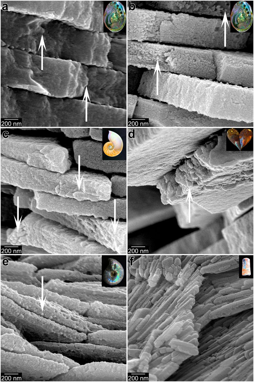
Finally, Schoepplera et al (2019) published in PNAS “Crystal growth kinetics as an architectural constraint on the evolution of molluscan shells” where they compare the process of ultrastructural morphogenesis of shells from 3 major molluscan classes: A bivalve Unio pictorum, a cephalopod Nautilus pompilius, and a gastropod Haliotis asinina. They demonstrate that the fabrication of these tissues is guided by the organisms by regulating the chemical and physical boundary conditions that control the growth kinetics of the mineral phase. This biomineralization concept is postulated to act as an architectural constraint on the evolution of molluscan shells by defining a morphospace of possible shell ultrastructures that is bounded by the thermodynamics and kinetics of crystal growth.
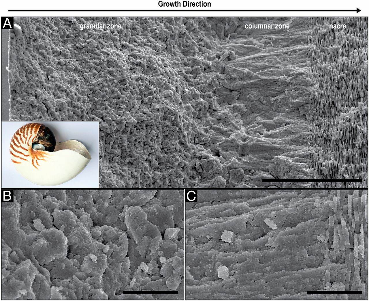
Structural analysis of the N. pompilius shell using electron microscopy (from Schoepplera et al 2019)
The study of biomineral formation is essential to our understanding of the most fundamental processes in evolution and organisms capability to adapt to environmental changes…so let’s keep investigating the “hard part of life”.