By Jack Wilkin.
One of the techniques used to examine fossils in the laboratory is photography with the aid of UV light. Ultraviolet light causes minerals in the fossils to fluoresce creating a clearer contrast between the fossil and the surrounding matrix. Ultraviolet photography is a cost-effective laboratory technique that can be readily applied to a wide range of strata types and taxonomic groups. It is quick to set-up and does not require specialised training.
History of UV light examination
UV light was first applied to palaeontology in the late 1920’s (Simpson, 1926; Miethe and Born, 1928). Most skeletal remains, minerals such as hydroxyapatite and phosphate, and sometimes slightly mineralized soft parts are fluorescent under ultraviolet light creating a contrast between skeletal parts and surrounding matrix (Hone et al., 2010). The Plattenkalks, or lithographic limestones, of Bavaria are a world-famous Konservate-Lagerstätten dating to the Tithonian Stage of the Late Jurassic (145-152 million years ago). The Plattenkalks represent a unique depositional environment with no comparable modern equivalents. The tests were conducted in the School of Earth and Environmental Sciences at the University of Portsmouth.
Methodology
Specimens for this study came from the Central Bavarian Mörnsheim and Solnhofen Formations. Fieldwork was conducted at Fossilienbesuchersteinbruch, near the village of Mühlheim, and Prinz-Max-Straße Quarry, near Eichstät, respectively. Specimens examined for this study included fishes, ammonites, and decapods (that is a group of crustaceans that include crabs, lobsters and shrimps). The south German Plattenkalks are very finely grained limestones that formed in isolated basins on the northern margin of the Tethys during the Upper Jurassic. The Plattenkalks are a Konservat-Lagerstätten that preserve a remarkable diversity of Jurassic flora and fauna from jellyfish to Archaeopteryx.
UV light is electromagnetic radiation with a wavelength between visible light and x-rays, in the range of 10 nm to 400 nm (Tischlinger and Arratia, 2013). Specimens should be carefully treated with a soft brush and sterile water to remove loose material and dust. Varnishing of specimens is not recommended as this may have affected the results. The optimum wavelength is to be tested experimentally as different specimens require different wavelengths to obtain the clearest possible results. Repositioning of the UV lamps is often needed to get the clearest results. It was in our experience during this study that there is no definitive distance to optimise the results, instead an experimental approach needs to be taken as each specimens has a optimum distance. The bulbs used are commercially available and can be brought cheaply. The wavelength used was 365nm though it is worth noting that different fossil groups fluoresce better under different wavelengths.
A long camera exposure is necessary to get detailed photographs. Both digital and analogue cameras can be used to capture UV images (Tischlinger & Arratia, 2013). The camera should be held in place using a stand to ensure it is steady during the exposure time. It is strongly advised to place smaller specimens in a box surrounded by black grains to stop flare and create a clearer contrast (Figure 1).

Figure 1: The techniques used to capture UV images. A: The positioning of the camera about the fossil being examined. B: During the image capture, the main lights were turned off. This is so the fluorescence of the specimen under UV would be more easily detected.
Health and Safety in the lab
Just as with the electromagnetic radiation emitted by the sun, artificial UV can be harmful. When exposed to bare skin, it can cause permeant skin damage leading to conditions such as malignant melanoma and other types of skin cancer so safety precautions must be taken (Tischlinger and Arratia, 2013).
Working in small enclosed spaces for extended periods with short-wave and mid-wave UV increases the local level of ozone. When working with such equipment, it is important to wear long-sleeved clothing and UV-blocking goggles. Even short-term exposure, and even indirect exposure, on unprotected skin or eyes, should be avoided (Tischlinger and Arratia, 2013). It is advised to take breaks every 10-30 minutes. These precautions should always be taken while working with UV. The safety equipment needed while conducting UV photography is cheaply and widely available.
Results of UV light
While UV techniques have proven to be useful in revealing sutures, other articulations, hidden bones, and the presence of soft tissues in fossil tetrapods and invertebrates it is rarely used in fish research (Tischlinger and Arratia, 2013). All the fossils recovered from the Plattenkalk and returned to the laboratory at the University of Portsmouth were placed under the UV light. All specimens, except for a few ammonites, showed autofluorescence under UV light. The UV light was able to show many of the fossil fishes more clearly allowing for skeletal elements to be identified (Figure 2), reveal previously unknown details, and even revealed previously unknown specimens (Figure 3).
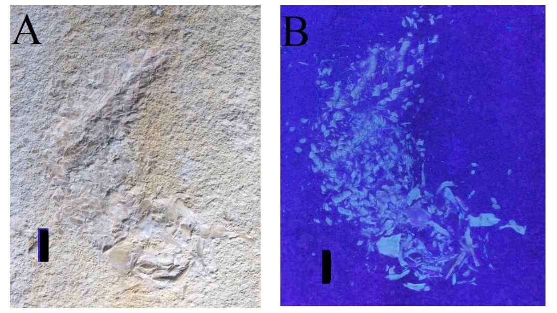
Figure 2: Skeleton of a Furo sp. Scale bar: 1cm A: Under natural light. B: Under UV-light. The UV reacts with biogenic minerals in the bones and scales causing fluorescence and reveals previously unseen details.
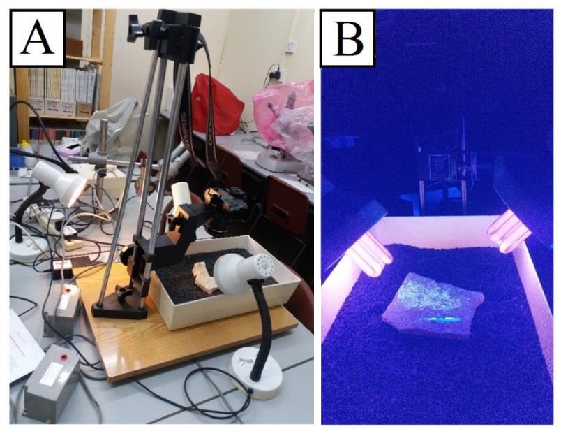
Figure 3: Image taken of the crustacean Eryon sp. This specimen was first identified in the field using a UV torch. A: Under normal light conditions, the fossil is hard to distinguish from the surrounding rock. B: When placed under UV the organic phosphate in the shell fluorescence’s and causes the fossil to become more visible.
The use of UV light can reveal critical information on the effects of diagenesis on a specimen. Fossils that have the least amount of diagenetic alteration show strong fluorescence under UV. This is because the elements responsible for florescence, phosphates, have not been replaced by other minerals during diagenesis and thus retain their original chemical signature.
Examination under UV light helps to identify uncertain anatomical features. For example, fish larvae a seldom identifiable in the Plattenkalks let alone collected for scientific study. When photographed under normal light the specimen is uninformative, but when placed under UV new previously hidden details can be observed providing important information on ontogenetic changes (Tischlinger and Arratia, 2013).
Each specimen responds differently to different wavelengths. Variables such as the wavelength used, exposure times, and positioning of the camera and UV source in relation to the subject must be adjusted for each specimen being photographed. It is not possible to apply a blanket approach to formations or even within a given horizon (Tischlinger and Arratia, 2013). The variables that may yield positive results from one specimen may not be appropriate for a specimen found in the layer above. Such variations are due to slight changes in the geochemical composition of the material being photographed and the environmental conditions in which it was formed.
UV light, along with low-angle oblique light, can also be used to identify forgeries. Controversy surrounding the authenticity of Archaeopteryx (Hoyle et al. 1985) were resolved with UV imaging. The use of UV light provided greater clarity proving that the skeletal elements were genuine and the cracks, presumed by Hoyle et al (1985) to be filled with cement, where infilled with minerals consistent with limestone diagenesis (Charig et al., 1986).
Conclusion
Ultraviolet light is an advantageous technique in Plattenkalk research. Its is cost effective and easy to setup requiring little to no specialised training. The use of ultraviolet light can be readily applied to different type of fossil locality but yields particularly good results in Plattenkalks.
ACKNOWLEDGEMENTS
Thanks go to Professor David Martill (School of Earth and Environmental Sciences, University of Portsmouth) for being my undergraduate project supervisor, Richard Hing (School of Earth and Environmental Sciences, University of Portsmouth) for helping with the UV photography and Roland Pöschl (Fossilienbesuchersteinbruch, Mühlheim) for allowing me to use his quarry.
REFERENCES
Charig, A.J., Greenaway, F., Milner, A.C., Walker, C.A., Whybrow, P.J. 1986. Archaeopteryx is not a forgery. Science 232(4750): 622-6.
Hone, D.W.E., Tischlinger, H., Xu, X., Zhang, F.C. 2010. The extent of the preserved feathers on the four-winged dinosaur Microraptor gui under ultraviolet light. PloS one. 5, 2, e9223. doi: 10.1371/journal.pone.0009223 PMID: 20169153.
Hoyle, F., Wickramasinghe, N.C., Watkins, R.S. 1985. Archaeopteryx. British Journal of Photography 132: 693-4.
Miethe, A., Born, A. 1928. Die Fluorographie von Fossilien. Paläontologische Zeitschrift 9, 343-356.
Simpson, G.C. 1926. Are Dromatherium and Microconodon mammals? Science 63, 548-549.
Tischlinger, H., Arratia, G. 2013. Ultraviolet light as a tool for investigating Mesozoic fishes, with a focus on the ichthyofauna of the Solnhofen archipelago. 549-560. In ARRATIA, G., SCHULTZE, H.P., WILSON, M.V.H (eds.). Mesozoic Fishes 5 – Global Diversity and Evolution. Verlag Dr. Friedrich Pfeil, Munich, 560 pp.
ABOUT THE AUTHOR
Jack Wilkin is currently a MByRes in geology student at the Camborne School of Mines (University of Exeter, UK) studying the isotopic geochemistry of belemnites for palaeoclimate studies. Contact: jack.wilkin@btinternet.com