Sorcha McMahon is a third year PhD student in the School of Earth Sciences at the University of Bristol. Sorcha is investigating how strange igneous rocks called carbonatites may have formed, using both natural samples and high-pressure experiments.

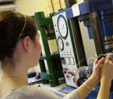
These back-scattered electron (BSE) images are a typical view of one of the high-pressure experiments that I run on the piston-cylinder apparatus, here in the BEEST labs at the University of Bristol. Such photographs are taken using the Scanning Electron Microscope (SEM), and are an essential stage in the analysis of run products as the different shades, textures and compositions are used to identify different mineral and melt phases.
The image on the right shows an entire capsule (a metal container that holds the powdered sample) and its contents after it has experienced conditions of 1375oC and 30 kbar (equivalent to ~100 km depth) for 24 hours. The AuPd capsule (an alloy that can withstand up to ~1400oC before melting at this pressure) appears brighter than the phases produced because this material has a higher atomic mass than the minerals (more information in Charly’s post).
The two images on the left show closer shots of the same experiment, labelled with the different minerals. In varying shades of grey; garnet, olivine, clinopyroxene (cpx) and orthopyroxene (opx) are typical minerals found in lherzolite (‘normal’ mantle) assemblages. As I am working in a synthetic carbonate-bearing system (CMAS-K2O-CO2), my run products contain an abundance of carbonate minerals, such as dolomite. At higher temperatures, melt may be observed, and is identified by its ‘streaky’ quenched texture.