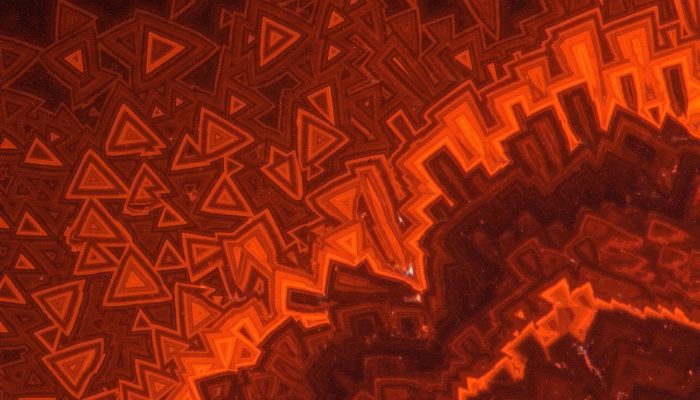
The truly insane photo above shows “Different generations of calcite cements in Late Miocene seep carbonates (Piedmont, Italy; cathodoluminescence microphotograph)”. It was taken by Marcello Natalicchio, University of Hamburg, Hamburg, Germania – Source.
This photo was taken using a cathodoluminescence microscope which is both a standalone tool or can be an adaptation to a scanning electron microscope. In cathodoluminescence microscopy an electron beam is fired at the sample which causes it to emit light (luminesce). The chemistry of the material being imaged determines the wavelength of the light emitted. Cathodoluminescence microscopy is a very useful tool for visualizing interior features such as fabrics, growth periods or other changes in the crystal interior that are not visible using traditional visible or polarized light microscopy. In the picture above the cathodoluminescence is being used to show the different stages of formation of a calcite cement.
In the geosciences cathodoluminescence microscopy is most frequently used to investigate the growth and deformation history of sedimentary carbonates, the interior structure of fossils, diagenetic processes, growth and dissolution of igneous and metamorphic minerals, and the growth of hydrothermal veins. The reason cathodoluminescence microscopy is so useful is that it allows the interior chemical variability of the sample to be imaged at the microscale without destroying the relationships that exist between the different stages of crystal growth.
I’m not counting Photo of the Week posts any more. The photo updates to the blog will now come whenever I feel like it. To be honest, I’m not sure how often I’ll feel like it. Although, now that daylight savings time has ended that may be more often since getting outside now involves enveloping myself in several layers of fabric. Also, daylight savings time is stupid, and I hate it.
Cheers
