For some years now, Mary Schweitzer and her team have been researching the idea that organic molecules can be preserved for millions of years, specifically within dinosaurs. They have used a plethora of chemical and biotechnological techniques to demonstrate that, within animals like Tyrannosaurus rex, it is possible to find the residue of structures such as blood vessels and even proteins. Naturally, her research has been met with a whole wad of stiff resistance from the scientific community, seemingly for no other reason than “We don’t like the sound of that..”. Scientific rigour ftw!
Recently, Schweitzer and her team released a new study digging deep into the geochemical basis for soft tissue preservation in dinosaurs. Using hardcore techniques like transmitted electron microscopy, electron energy-loss spectroscopy, micro-X-ray diffraction and Fe micro-X-ray absorption near-edge structure (methods that have been borrowed from geochemists and environmental sciences), it is possible to look at the ‘chemical ghosts’, a term coined by Phil Manning, that are associated with fossil remains.
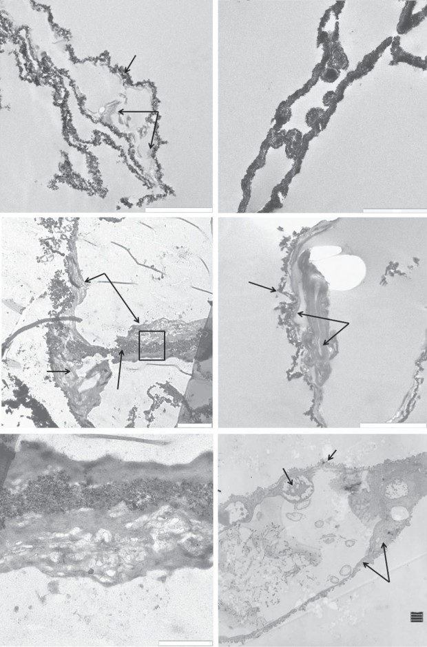
Transmitted electron microscopy images of (a,c,e) T. rex vessels, (b,d ) B. canadensis vessels and (f ) ostrich vessels. T. rex vessels show iron particles infiltrating an ‘organic’ layer . B. canadensis vessels are more completely infiltrated with iron (b), but in some views (d ), an organic layer is still visible. Scale bar, 2 mm for (a–d), 1 mm for (e,f ) (Schweitzer et al., 2014)
For me, this is one of the greatest steps in recent palaeontology – no longer do we just have bones, but we have other soft tissues like feathers, skin, and internal structures, adding a whole new bio-chemical dimension to how we perceive fossils. Of course, this opens up a whole new wealth of knowledge to be uncovered about extinct animals, their physiologies, and their evolutionary roles.
The previous lines of evidence supporting the cellular-level preservation of soft tissues (see bullet points below) all require a mechanism whereby preservation and mineralisation outpaces the decay of soft tissues. This is the point where Olay and Chanel get interested in Palaeontology. Schweitzer’s new evidence points towards a type of ‘tissue fixation’ that leads to the chemical preservation of molecular organic materials. The main thrust being that iron can contribute towards the exceptional preservation of soft tissues and molecules after death.
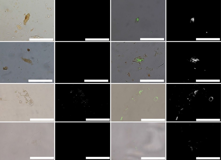
Overlay and fluorescence microscopy images of dinosaur ‘osteocytes’ and vessels after exposure to polyclonal antibodies. Scale bar is 50 mm.
In science, controls are key. These are like standardisation tests that demonstrate your analyses are giving results that either differ or are the same as some known result. For example, one of interest these days would be the background chemicals in aquifers that have to be known before you can start attributing contaminants to fracking fluids. The same is in palaeo – when running analyses on fossilised organisms, you have to know that these tests are valid on modern ones too, so they show the same thing. So not only did Schweitzer and her team perform their array of tests on Tyrannosaurus rex and the hadrosaur Brachylophosaurus canadensis, but also an ostrich just to demonstrate their applicability and validity in modern analogues (ostriches are one of the most ‘primitive’, or basal, birds alive today).
In the ostrich, iron accumulations formed quickly on certain parts of the ostrich tissue, and it was these elements that resisted decay for the longest, showing that iron can enhance preservation in modern animals, specifically extant dinosaurs. In both T. rex and B. canadensis, the same is found – iron is found strongly associated with structures like blood vessels.
In preserved osteocytes, a star-shaped cell found in bones, the same intimate relationship between iron and organic remains is found too in both T. rex and B. canadensis. This provides additional evidence that these are actually the exceptional remains of soft tissues, and not some external contaminant of some sort. Each additional analysis the team conducted continued to show the same signal – that iron, particularly in an oxidised stated, is strongly associated with cellular or vessel like structures, identical to the relationship found in ostriches.
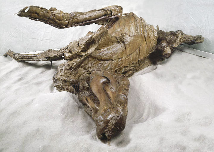
One of the infamous dinosaur mummies, a hadrosaur entombed in the flesh for 70 million years. (source)
This is a pretty damn compelling case if you ask me (I know you didn’t, but it still is). It’s a strong case for cellular-level preservation in dinosaurs, something that 10 years ago would have been laughed out of the room, and still is by many. It goes another step too – while iron and oxygen are both crucial elements for animals during life, it seems now their role is taken to the grave and they play a critical role in the preservation of biological remains, even after tens of millions of years.
Together, this new study adds to a host of lines of evidence that point towards mineralisation of soft tissues at a teeny microscopic scale within dinosaur bone, including:
- Reaction of antibodies through binding with chemical extracts and within the bone (the biotech part) – these are not reactive to microbial biofilms, ruling that out;
- Peptide sequence data from the preserved proteins, not found in microbes either;
- The finding of nuclear chromosomal proteins called histomes, that based on their locality and amino acid sequence data indicate a biological origin, again, not from microbes.
More importantly, how much have palaeontologists missed by not considering these potential high levels of preservation in dinosaurs? And how much is there still left out there to be found at these exquisite levels of detail? It’s cool though – palaeontology seems to be diverging into two major pathways: the analysis of large datasets to look at really big global trends through time, for example in biodiversity; and the microscopic, by exploring the chemical ghosts that fossils have left behind for us to trace.
Reference:
Schweitzer, M. H. et al. (2014) A role for iron and oxygen chemistry in preserving soft tissues, cells and molecules from deep time, Proceedings of the Royal Society B, 281, 20132741 (link) (open access version)
David Mennear
Excellent post! I had heard of Schweitzer’s work before and always wondered why it was almost put to one side. Never seen the hadrosaur ‘mummy’ before, thrilling 😀
Jon Tennant
Plenty of dino mummies out there! Most famous is one called ‘Dakota’ – there was a documentary on it a while back, worth trying to track down
dwbapst
I still find the graptolites more amazing.
dx.doi.org/10.1038%2f237443a0
Jon Tennant
Wow, that’s pretty awesome! The fossil record is full of nice surprises 🙂
Timothy Huang
Regarding to organic remains preserved in dinosaur, actually in many fossils, please see Robert Reisz and my Nature cover paper, Nature April 11, 2013, p 210-214
Embryology of Early Jurassic dinosaur from China with evidence of preserved organic remains
By the way, this cover photo was selected by NPG as one of the annual photos. And our paper was again reported by the Russian newspaper (http://kompravda.eu/daily/26177.5/3066776/) as #4 of the most outstanding scientific achievement of 2013.
Pingback: Morsels For The Mind – 28/02/2014 › Six Incredible Things Before Breakfast
Nathaniel Latham
I’m reading this and the only thing I’m seeing is Jurassic Park? Jurassic Park, Jurassic Park…. Jurassic park!
Jon Tennant
Ha, it’s probably not as absurd a thing as it was 10 years ago..
Pingback: The Geek’s Reading List – Week of March 7th 2014 | thegeeksreadinglist
Pingback: Chemical ghosts of dinosaurs may help reveal new secrets - Technology Org
Pingback: CEH: Rescuing Dinosaur Soft Tissue from the Ravages of Time
Pingback: Rescuing Dinosaur Soft Tissue from the Ravages of Time - Creation RevolutionCreation Revolution
ashley haworth-roberts
A dogmatic young earth creationist (with an astronomy background) is accusing Jon of bad practice:
http://crev.info/2014/03/dinosaur-soft-tissue/
Pingback: How Gaps Are Filled In Evolution | New Discoveries & Comments About Creationism